There is something exciting about thinking about Masiakasaurus knopfleri. It’s not just the name’s tip of the hat to Dire Straights’ lead guitar and frontman, Mark Knopfler, or the becoming-more-prevalent use of local language to name the animal (masiaka means “vicious” in Malagasy, a play on which occurs in the title, and the gorgoylesque visage of which is fitting as a blog post for this All Hallows Eve, aka, Halloween).
Matt Carrano and colleagues, in 2001, reported a new taxon from a diverse array of specimens including multiple different individuals (based on overlapping material), selecting as the holotype a unique dentary (UA 8680). This jaw is unusual, for the first demonstrating in a dinosaur downward-curving arc of the dentition, where the dentition became larger and longer towards the front, resulting in anteriorly projecting front teeth.
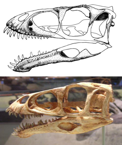
Skull reconstructions of Masiakasaurus knopfleri, top: from Carrano et al. (2011), bottom: from Wikimedia Commons.
This feature is unknown in any other known theropod dinosaur, and so it was presumed unique enough to grant the animal a new name. Numerous shed teeth from the sites where the material was collected, as well as an array of addtional dentaries (Carrano et al., 2011, report 8 total dentaries known), show that the morphology is consistent across a range of sizes, and a large, complete dentary with in situ teeth show the form of the slightly procumbent to strongly procumbent or nearly horizontal anterior teeth. This makes the jaw pretty weird.
It doesn’t take any spark of imagination to think this animal was doing something with that jaw different from what other theropods might be doing with theirs. Most theropod jaws have teeth in the front largely vertical, and even if they were slightly forward pointed (as in Coelophysis bauri), there are none to this degree in theropods with one very interesting exception: Epidexipteryx hui, a scansoriopterygid with extremely large procumbent teeth (as I noted here, calling it a “nitpicker”). In Epidexipteryx hui, the lower and upper teeth are restricted to a small, anterior section of the jaws, clustered together and largest in the front; the teeth are generally arrayed at the same angle, projecting around 45 degrees from an implied horizontal (ventral margin of the maxilla); and the teeth are slightly recurved, with higher curvature in the posteriormost, and shortest, teeth. In Masiakasaurus knopfleri, these teeth are spaced a bit more, and only the front teeth are so angled, but the generally morphology seems to be the same.

Masiakasaurus knopfleri, Daemonosaurus chauliodus, and Epidexypteryx hui. From Carrano et al. (2011), Sues et al. (2011), and by me, respectively.
But similar jaws are known in non-dinosaurs, especially in archosauromorphans like riojasuchids, proterosuchids, but also many “notosuchians,” such as the “dog crocs” Araripesuchus. There, the rostrum is downturned from the line of the maxillary teeth, and the premaxillary teeth are somewhat inflected posteriorly. This is also true in pholidosaurid eucrocodilians, such as Sarcosuchus imperator (de Broin & Taquet, 1966). The jaws are also relatively shorter by a distinct measure than in animals with more precise rostral occlusion, as in the gharial, and the rostral mandibular teeth tend to be oriented anteriorly, often in a radial (or circular) array. Strongly decurved rostra and much shorter mandibles than upper jaws with anteriorly oriented teeth are also the rule in rodentians and lagomorphan mammals, where the occlusion of the incisors occur at a distinct lower angle than the more posterior teeth. There is often a significant diastema between the incisors and the premolars/molars in rodents, fossil or otherwise. Horses, especially Equiidae, also have a decurved rostrum, posteriorly oriented upper and anteriorly oriented lower teeth, and this is readily also apparent in many other perissodactyle clades, including rhinoceratids.
All of these features result in a somewhat pronounced overbite, and the functional effects of this are generally related to acquisition of food at the very front of the jaws. The effect of the paired angulation — where present — of dentition may be functional: the orientation of the upper dentition may reflect line of action in some teeth, especially when straight, while in curved teeth reflects loading resistances (as in rodents) to proximodistal compression, useful when you are applying high pressures to the tips of a few teeth compared to the area of jaw being affected. Otherwise, evenly spaced dentition, lacking a diastema, would result in less exaggeratedly oversized teeth in some taxa, as in gharial crocodilians, but endorse it in others, as in spinosaurid theropod dinosaurs.
The rear-ward orientation of the upper teeth and the overbite enables a downward motion with the skull instead of a jaw-only or equally paired jaw/mandible acquisition, reduced need for leverage over the food as in rodents/lagomorphans, and in predators enabling the lunging strike, concentrating more force in the rostrum than in predators where the rostral and midline/posterior teeth are more even in size.
This latter effect applies directly to some dinosaurs, such as hadrosauroids (which lack teeth in the anterior rostrum, but have the downturned rostrum and overbite in most known taxa), but even more interestingly in “coelophysoid” Theropoda. In these taxa, there is even a distinct gap between the premaxillary and maxillary teeth, and the premaxilly is offset from the lateral margin of the maxilla, so that the posterior margin is somewhat medial to the maxilla, and thus results in a slight “pinch.” The pinch is copied in spinosaurids like Suchomimus tenerensis (or, Baryonyx tenerensis, or Cristatusaurus [Baryonyx?] lapparenti, or whatever it “should” be called), but as in the gap in heterodontosaurids, this pinch does not displace the posterior premaxilla medial to the maxilla, mearly dorsal to them, but still narrow. Sakamoto (2010) discussed this in relation to estimating bite force efficiency along the jaw, where in Coelophysis bauri and Dilophosaurus wetherilli the premaxilla is oriented rostroventral and the premaxillary teeth angle caudally. In the mandible, the rostral dentition even includes a radial array of the rostralmost several teeth.
There should similarly be large ventroflexor muscles of the head-neck region, indicated by large processes of the proximal cervical vertebrae and the basioccipital and palatal region of the skull not connected to jaw adduction. However, in Masiakasaurus knopfleri, these regions of the skull remain largely undescribed; the ventral braincase is described for one specimen (Carrano et al., 2011; FMNH PR 2457), in which the basioccipital tubera, attachment sites for some head-flexors such as m. rectus capitis, are short, rounded, and not distinctly separated from the occipital condyle, unlike the condition in Baryonyx walkeri (Charig & Milner, 1997). A well-developed ventroflexor region of the head-neck muscles should correspond to primary processing of the jaw by application of the rostrum, rather than the mandible, or by orthal biting with puncture-pull methods of dismemberment/processing (Hertel, 1995), something that compares well with the distinction between birds with large-hooked beaks (falconiform and strigiform birds) and those without. These birds also exhibit distinct overbites and a mandible curved rostroventrally, matching the curve of the upper jaw. It should be evidence, then, that the curvature of the upper and lower jaws might then be expected to coincide with one another, rather than depart strongly. The margin of the jaw formed by the teeth or the mandible itself in the absence of teeth should compare strongly to the toothed or beaked-margin in the other, regardless of the overlap of the former by the latter (as in falconiforms, strigiforms and psittaciforms; but also procellariforms).
The Face of an Angel
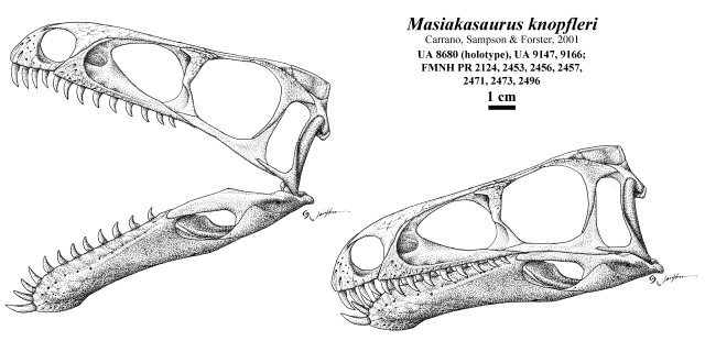
A reconstruction is offered below, of the rather unusual skull anatomy of Masiakasaurus knopfleri.
[Before anyone says anything, I’m not wholly satisfied with this reconstruction, and will likely change some features automatically in the future. Such are the vagaries of reconstruction from a large array of differently-sized animals and low confidence of “adult morphology”.]
This skull is broadly restored after the specimens indicated, but rather than simply drop the various specimens side by side, I’ve tried to scale them against one another using the intact skulls of Coelophysis bauri, “Megapnosaurus” kayentakatae [possibly “Kayentavenator” kayentakatae], and Limusaurus inextricabilis. This has included, but is not limited to, making the dentary comprise a large portion of the mandibular length, the rounding of the orbit with small postorbital region, suggesting that the premaxilla preserves maybe 1/2 its natural length, and scaling the maxilla up to the point where it comprises a similar ratio of total and ventral skull length, and matching the sizes of the maxillary and dentary dentition. As can be compared above, this skull provides a larger snout-to-skull comparison than Carrano et al. (2011), and may imply an overall longer skull with less-rounded orbit than has been suggested by art since it’s description.

Skull reconstructions of Masiakasaurus knopfleri Sampson et al., 2001. A and B modified after Carrano et al., 2011; C from Andrea Cau’s blog (link is below), D, my reconstruction; E, dentaries, based on – E1, FMNH PR 2471 after Carrano et al., 2002 and E2, UA 8680, holotype, after Carrano et al., 2011. Scale bars in E equal 10 mm, blue in E represents missing material.
C, above, from Andrea Cau, who responded with this reconstruction following Mickey Mortimer’s comment:
I’m not impressed by the cranial reconstruction, which doesn’t match the preserved morphology. Even ignoring the misleading appearence of completeness for the posterior premaxilla and dorsal lacrimal, the maxilla below the antorbital fossa is too narrow, the lateral lacrimal flange incorrectly overlaps the antorbital fenestra, the postorbital is too robust, the frontal margin of the orbit is too short, etc..
Direct comparison reveals not only some difficulties with the reconstructions, but that the maxilla makes an ill fit (it is too small for skulls in the reconstructions by far, for various reasons) and that this influences how these reconstructions come about. First, and most importantly, the maxilla is represented by an individual smaller than that of the well-preserved dentary FMNH PR 2471, and suggested by the shorter length of the alveolar margin and its incomplete nature (as the represented maxilla, FMNH PR 2124, preserves only 6 of what were likely more dental alveoli) but also by the problems when scaling up to “match” the dentary: the dental alveoli should normally be the same size, but with maxillary alveoli on the larger side and with either an even number of maxillary to dentary teeth, or a greater number, per a given length. The reconstructions go in the opposite direction.
Second, my reconstruction infers growth toward the upper end of the size of elements, but doesn’t pick one element to be the best or “right” size, due to the lack of clear ontogenetic age in the various fragments. This influences the relative shapes and positions of bones: I increased the distance between the lacrimal/prefrontal and the postorbital, despite suggestion that the orbital rims of each bearing “ornamented” features that might surround the orbit. There is no reason to assume that the orbital rim is circular in this reason, despite this, although it is possible. Similarly, I had to elongate the frontals a bit to coincide with interorbital and preorbital elongation.
Third, I did opt to go with the holotype (UA 8680) for the shape of the rostral end of the dentary, and the orientation of the mesialmost dentition. In the larger specimen, the rostral dentary is more angular; one can assume that the larger specimen represents an older morphology consistent with ontogeny, however I am unaware of ontogenetic trends to suggest otherwise. Should I have more reason to correct this, I will. One of the problems with this is the concern over the shape of the dentary in lateral view, especially given how the original descriptive argument was that the dentaries are not perfectly parasagittal, but that the alveolar margins are oriented labially, while the gular margin was oriented lingually somewhat. How this affects the lateral aspect of the dentary depends on the degree of splay: an angle of 45° can result in a vertical shortening by as little as 5%, but also an alteration of the shapes of the bone. Now, the reconstruction takes UA 8680 (holotype of the species) at face value and extrapolates it by size with some features added in from FMNH PR 2471; a possible revision, pulling out the former specimen for the latter, would not result in much distinction, as I am continuing to argue that the dentary may be undersized for an adult animal, which my reconstruction approximates.
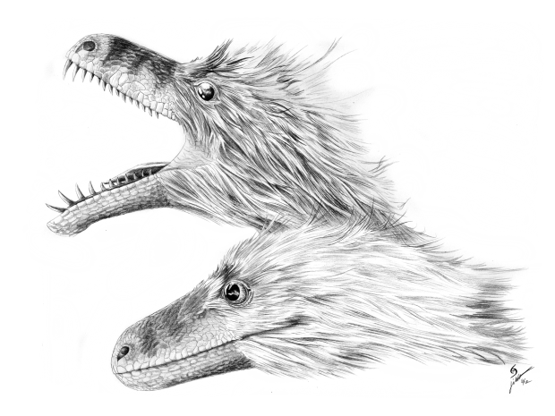
Previously, I also offered this life reconstruction of a theropod, and asked viewers to guess it. Many viewers suggested something pretty basal, and many guesses were focused around basal Theropoda, or even non theropodan saurischians, or some tetanuran theropods. No one guessed the reconstruction might be that most weird-toothed of theropods, Masiakasaurus knopfleri. The reconstruction was only half of an illustration, and the other (had I shown it) would have given the game away. Here it is in full:
I was being quite liberal with the soft-tissue, and the shapes of the skull bones are almost entirely obscured due to this amount of flesh. Moreover, a good deal of flesh is needed to cover all of the teeth, and so the extraoral tissues are rather large. I imagined, as I reconstructed this, the head of a monitor lizard, and the complete inability to define bone shapes from the shapes of the fleshy head.
Choppers
Masiakasaurus knopfleri exhibits some interesting teeth (Sampson et al., 2001; Caranno et al., 2002), in which the rostral dentition of the mandible are elongated, semi-conical, and in the distalmost crowns have carinae twisted labiolingually rather than mesiodistally; but in more distal crowns, the teeth are shorter, strongly labiolingually compressed, and the carinae are oriented in “typical” mesiodistal fashion, and they are all somewhat oriented rostrally, mesially inclined at the base (a morphology consistent even with isolated teeth from Brazil: Lindoso et al., 2012).
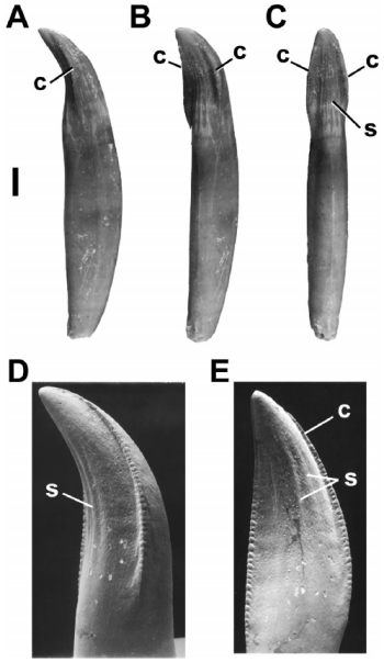
Mesial mandibular teeth (FMNH PR 2180, 2182, and 2200 ) of Masiakasaurus knopfleri, from Carrano et al., 2002.
At least one consideration on tooth functionality, raised by Fowler et al. (2011) ,was that the orientation of denticles indicate orientation of tooth action, but it also suggests that orientation of carinae affect tooth action. In Masiakasaurus knopfleri, the denticles are oriented apically, although it is not clear if they are hooked (Carrano et al., 2002, 2011, do not describe the denticle morphology in detail), while carina orientation changes along the tooth row; rather than follow the curvature of the jaw margin, as the carinae do in “typical” theropods (e.g., tyrannosauroids), the teeth become somewhat splayed and the carinae rotate around the tooth long axis as the teeth become more and more horizontally oriented. This results in the mesialmost dentary teeth having a labiolingually-oriented carinal “blade” in the rostralmost pair of teeth, which were almost certainly adjacent, while the more distal teeth gradually become more “normal”. This suggests that the rostral dentition were affecting their food in a fundamentally different manner than might be expected if, instead, the teeth were interlocking in a gharial-like rostral array. This calls into question the inference that Masiakasaurus knopfleri may have been a fish-eater of some acclaim (Sampson et al., 2001, and virtually all later references). Interestingly, despite apparent interlocking arrangements of the dentition (Sampson et al., 2001), the teeth lack wear facets, implying despite the close spacing rostrally, the teeth probably did not interlock, but rather like Crocodilidae, Araripesuchus and virtually all Theropoda, the lower dentition was probably lingual to the upper dentition. The labial splay of the dentition may have been exaggerated.
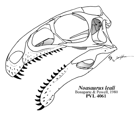
Indeed, this aspect suggests that, while rostral prehension is present, a complex oral processing suite was also present, with distinct segregation of functional utility of the rostral and distal components of the dentition. Following reconstruction of the skull of Masiakasaurus knopfleri, and specifically the implication that the upper and lower jaws should have comparable curvature, one can apply this model to Noasaurus leali (Bonaparte & Powell, 1980), and it suggests that the mandible is even more curved than in Masiakasaurus knopfleri, but also that the rostrum was further flexed and the mesialmost dentition further caudally facing.
Previously: Do they dream of algal sheep? Triassic palate mashers.
Up next: We’ll see.
Bonaparte, J.F. & Powell, J.E., 1980. A continental assemblage of tetrapods from the Upper Cretaceous beds of El Brete, northwestern Argentina (Sauropoda, Coelurosauria, Carnosauria, Carnosauria, Aves). Mémoires Societe Geologie France 139:19-28.
Carrano, M. T., Sampson, S. D. & Forster, C. A. 2002. The osteology of Masiakasaurus knopfleri, a small abelisauroid (Dinosauria: Theropoda) from the Late Cretaceous of Madagascar. Journal of Vertebrate Paleontology 22(3):510-534.
Carrano, M. T., Loewen, M. A. & Sertic, J. J. W. 2011. New materials of Masiakasaurus knopfleri Sampson, Carrano, and Forster, 2001, and implications for the morphology of the Noasauridae (Theropoda: Ceratosauria). Smithsonian Contributions to Paleobiology 95:1-53.
Charig, A. J. & Milner, A. C. 1997. Baryonyx walkeri, a fish-eating dinosaur from the Wealden of Surrey. Bulletin of the Natural History Museum of London 53:11-70.
de Broin, F. & Taquet, P. 1966. Découverte d’un Crocodilien nouveau dans le Crétacé inférieur du Sahara [Discovery of a new crocodilian from the Lower Cretaceous of the Sahara]. Comptes Rendus Hebdomadaires des Séances del Academie des Sciences, D: Sciences Naturelles 262:2326-2329.
Hertel, F. 1995. Ecomorphological indicators of feeding behavior in recent and fossil raptors. The Auk 112:890–903.
Lindoso, R. M., Medeiros, M. A., Carvalho, I. de S. & Marinho, T. da S. 2012. Masiakasaurus-like theropod teeth from the Alcântara Formation, São Luís Basin (Cenomanian), northeastern Brazil. Cretaceous Research 36:119-124.
Fowler, D. W., Freedman, E. A., Scannella, J. B. & Kambic, R. E. 2011. The predatory ecology of Deinonychus and the origin of flapping in birds. PLoS ONE 6(12):e28964.
Sakamoto, M. 2010. Jaw biomechanics and the evolution of biting performance in theropod dinosaurs. Proceedings of the Royal Society of London, B: Biological Sciences 277:3327-3333. [PDF]
Sampson, S. D., Carrano, M. T. & Forster, C. A. 2001. A bizarre new predatory dinosaur from Madagascar. Nature 409:504-506.
Sues, H.-D., Nesbitt, S. J., Berman, D. S. & Henrici, A. C. 2011. A late-surviving basal theropod dinosaur from the latest Triassic of North America. Proceedings of the Royal Society of London, B: Biological Sciences 278:3459–3464.
Up Next: Do they dream of algal sheep? Triassic palate mashers.




Your reconstruction seems to inherit the same mistakes Carrano et al. made- maxilla too narrow below antorbital fossa, lateral lacrimal flange extends anteriorly to level of antorbital fenesta, etc.. Almost as if you used their reconstruction as a reference instead of the photographs of each element.
“No one guessed the reconstruction might be that most weird-toothed of theropods, Masiakasaurus knopfleri.”
Because it doesn’t fit over the skull, even your reconstruction of it. Since both have closed mouths, we can use the snout height to scale them, and we come up with this- http://i61.photobucket.com/albums/h64/therizinosaurus/MasiakaJaime.jpg . Which would be the only way to avoid your lack of a ventrally concave dentary- making the straight ventral edge throat tissue. Though that superposition is clearly not what you had in mind, as the eyeball would be in the postorbital and the maxillary lips would be huge (though not the premaxillary lips). No doubt you intended a match closer to this- http://i61.photobucket.com/albums/h64/therizinosaurus/MasiakaJaime2.jpg . Now the eye is in the right place and the lips are reasonable, but as I said previously, the ventral edge of the mandible isn’t properly concave (shown in red and blue lines). You can even tell you didn’t intend extensive throat tissue because you did such a good job matching the scalation patterns that we can scale your reconstructions (http://i61.photobucket.com/albums/h64/therizinosaurus/MasiakaJaime3.jpg) and see almost all of the mandible is visible when the mouth is closed and the only difference is the close-mouthed one lacks a downturned synphysis, which was of course something that couldn’t flex up or down in the live animal.
You’re an excellent artist, Jaime, and your Dr. Jekyll and Mr. Hyde theme is fun, but the truth is Masiakasaurus would have still looked different from a dromaeosaurid or basal coelurosaur even with its mouth closed.
I’m not sure what the issue with the maxilla is: The maxilla is restored close to the same shape as the preserved bone, with some inferences drawn from Noasaurus leali for the missing lacrimal/maxilla contact dorsal to the antorbital fenestra. The lacrimal itself isn’t that much of a problem, as I see it; while illustrated largely from Carrano et al., I followed the bone more closely than you might think but rotated it slightly medially on its rostral end, and as the lateral margin of the lacrimal already appears to approach if not meet the medial margin in lateral view, these should “meet” or the latter be exceeded by the former in rostral extent in life … not to mention a suggestion that age should emphasize the ornamentation of the lateral surface and expand it. Yes, a broad assumption of some of the shapes of Carrano et al’s reconstruction was involved, but rather than assume they were wrong and rather than assume that I wasn’t dealing with more data than they were (as they haven’t illustrated all cranial bones presented), I made the decision to use their reconstruction as a general model and fitted the real bones to them, with some assumptions of extending ontogeny or scaling up. I am not exactly dealing with perfect information here, nor am I taking bone shapes at face value as much as you are.
As I explained to you before about the ventral margin of the jaw, you seem to be thinking that no manner of soft tissue should cause the jaw’s lateral shape, or the large, numerous foramina to have anything to do with any amount of additional soft tissue, but instead more of a shrink-wrap with “lips”, a la Greg Paul. That is quite certainly what I am trying to avoid! The gular margin of the dentary SHOULDN’T be visible due to the fats, skin, and muscles of the gular tissues, and despite the concave gular margin of the dentary, the symphyseal region with its distinct lateral groove and numerous, large foramina, it seems more reasonable to me to imply a more gradual curve of tissues in lateral aspect from rostrum to jaw hinge, and obscure the rostral end of the dentary, which shouldn’t appear as if shrink-wrapped in life.
Finally, the consequence of the life appearance is unintentional, save for the use of hefty filamentous extraintegumental structures; I think I have a good case for the integument thickness and filaments, although that is a resonable extrapolation through phylogenetic bracketing from basal ornithischians and theropods. Despite this, a good number of people suggested even more basal theropods or saurischians, so the integument itself isn’t necessarily a cause for confusion. The squarish, “boxy” skull in profile may be a factor of constraint across small theropod dinosaurs, and not something that arises out of bias. While coelophysoids have shallow, inconspicuous premaxillae, which help slope the snout downward and provide a triangular profile, abelisaurians seem to have deeper premaxillae: I am simply extending that premise to Masiakasaurus knopfleri and other noasaurids.